Pages

Thursday, September 15, 2011

The Functional Frog and What Can Go Wrong

Who on earth looked at the bottom of a horse's hoof and thought "I will call that part... THE FROG!".

Anyway.

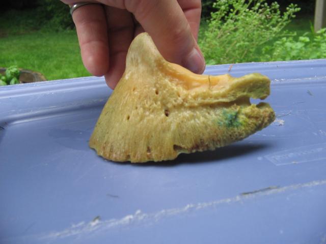
A near perfect frog, from a hind hoof.
The frog's function is to be an essential part of the landing gear at the back of the foot. It acts as a shock absorber along with the digital cushion. Also, when the hoof hits the ground, the frog compresses and squeezes blood back out of the foot and up the limb again.*Edit 29.10.11: there is some research that suggests that when the hoof is loaded, the dropped frog and sole actually create a vacuum ad suck blood into the hoof, rather than pushing it out! Must investigate this further... Ok, carry on!* I like to say that a horse has five hearts - one in his chest and one on the underside of each of his feet. :)

Correct movement = heel first landings = the force of the hoof hitting the ground being dissipated throughout the structures designed to do just that, limiting concussion.

The frog should look plump, open, flat and healthy. It should be wide at the heels. The central sulcus should look like someone has pressed their thumb into pliable putty, not look like a deep crevice. It should be free of thrush, and have passive contact with the ground in the stationary hoof. On soft ground (where the hoof would sink into the ground when loaded) the frog should be further away from the ground than on hard ground, where the frog is normally at heel height

The frog should take up 2/3 of the hoof


The frog should barely be trimmed at all. The only trimming a frog would need in most circumstances is to open up any areas that thrush would be hiding under.

Here is a trim I did on Gracie. Compare the frog before (left) to after I finished the trim. All I have removed is anything that was harboring thrush but did not remove anything she would need. (Ignore the terribly uneven heels - this was fixed after photo was taken!)

I found this on the internet - the whole frog needing to come off as there was a severe thrush infection under there. Thrush is painful! This horse would need boots and frog stimulating pads (depending on how comfy he was in them) on 24/7 til the frog calloused and came back to life I would imagine.

The frog should NEVER be trimmed like this, no matter how pretty that looks!
If the frog is compromised in any way (thrush and over trimming being the most common), this leads to flat or toe first landings. If the hoof is trimmed (or shod) in such a way that the frog is taken out of it's secondary weight bearing duties, then the hoof will contract up, the central sulcus will form a deep crack and provide a nice comfy house for thrush to thrive, and the frog will no longer be fully functional (if at all depending on the amount of contraction).

Frog has become too passive due to thrush infection (looks like it is clearing up though)

Severely contracted hoof and frog.
Top is a fairly normal hoof with frog having passive ground contact. Bottom is a contracted hoof with the frog suspended out of contact with the ground unless the ground was very soft.

So consider the frog when trimming - leave it alone unless there is thrush present, then take the bare minimum you can. Keep it clean, and in passive contact with the ground. Most of all, trim for heel first landings! If a horse is landing heel first, the frog will pretty much look after itself.

Thursday, September 8, 2011

Canker?!

Here is Australia it is cheap and easy to keep horses living outside in yards and/or paddocks 24/7. Most of the country has relatively mild winters (only small pockets of the country actually get snow) and a few good waterproof rugs is generally all you need for winter.

So, I have not been exposed to the delights and pitfalls of the stabled horse. There is a lot about managing the stabled horse that I have no clue about or have never heard of. One of those things is Equine Canker.

I was googling for some pictures of frogs for an upcoming post and came across this canker business. I thought canker was just an old English word for cancer. And apparently that is sort of what it is - hoof cancer. The pictures I saw were grotesque. I'll post a few at the end of this post.

Here is a link to an Aussie PDF info page about canker:
http://www.barehoofcare.com/fact%20sheets/equine_canker.pdf

The short of it is that canker is an eventual result of infected feetsies standing around in excrement for too long. It is an over growth of hoof tissues and apparently smells pretty disgusting due to the overactive cells getting infected with all sorts of vile and disgusting microbes. Yummo.

It is treatable but is degenerative and needs to be sorted out pretty early apparently.

I have never even heard of this. So there you go.

Sunday, September 4, 2011

Notes on the Pedal Bone - *warning cadaver pictures*

(Please be aware that there will be some pretty gross pictures of hoof disections in this post - please try to check them out though, the knowledge gained is invaluable!)


An x-ray of a normal hoof. 'A' indicates P3. (B is the navicular bone, c is P2 or the short pastern bone, and D is P1 or the long pastern bone).
A cross section of a cadaver leg.
 
This is an awesome digital image of P3 in relation to the lateral cartilages (light blue) and tendons.

Pedal Bone, Coffin Bone, P3, Distal Phalanx - this bone goes by a few different names. I like to refer to it as P3 as it is quicker to type and say!

P3 is the primary bone in the horse's hoof. To me, it looks like a little miniature version of a hoof - the outer hoof should match the bone on the inside. A bigger, flatter-soled P3 will show a flatter soled larger hoof. A very concave P3 bone will show as a hoof with magnificent concavity. There are soft tissues and blood vessels attached to P3 including the soft laminae that attach the bone to the hoof capsule.

Hoof with capsule removed showing the solar corium (the blood filled pad separating the sole and lower surface of P3), the soft laminae and the coronary corium from where the hoof horn grows.
Look at the solar surface! The sole and frog corium (which is just a fancy name for soft tissues if you ask me) looks like a play-doh cast of the hoof! This shows why it is important to have sufficient sole depth.

I did some googling and there are some VERY interesting pictures of P3 bones out there!

Various P3 bones of front feet from the top. The one top left has significant damage probably from an abscess or infection of some kind.

Same bones as the picture above but viewed as if holding the hoof in your hand to be picked. Note the different soles and how they differ in concavity.

As viewed from the side.

Here is P3 in the hoof capsule
And again on a shod hoof. Here it shows really well the concept that the collateral grooves (the valleys beside the frog) are a good indicator of sole depth - the shallower they are, the less sole you have.

You will hear about P3 being talked about in relation to rotation and balance, particularly with cases of laminitis.

Anterior to posterior balance refers to the angle of P3 in relation to the ground surface. The accepted norm is 3-5 degrees rotation when viewed from the side. This is to allow some movement of P3 and the structures in the back half of the foot to 'sink' when the hoof is loaded and expands (that is why it is important to trim in quarter scoops to follow the live sole plane).

The red lines show the amount of rotation - imagine this hoof being loaded when taking a step - the angle made by the two red lines would close when loaded to absorb impact.


Laminitis is as we all know when the laminitic structures loose their connection for a plethora of reasons and P3 can and often does rotate.

Here you can see the tip of P3 making the sole bulge just in front of the frog. Your brain should be screaming a warning at you if you ever pick up a hoof and see this! This is why a laminitic horse uses the infamous 'laminitis' stance - where a horse throws his hooves out in front of him and loads his heels to take the pressure off  P3 and the excruciatingly painful laminae)

An x-ray of a laminitic foot. See how P3 has rotated, the hoof wall has ripped away from the front surface of P3? See how the foot would bulge?


The best way to see without an x-ray machine if the horse has an ideal or acceptable amount of rotation is to draw a line straight through the pastern and hoof - the angle of the hoof capsule should closely match that of the pastern.

Here is Gracie back in March this year - you can see the angle of the pastern vs the hoof is broken forward - the tip of P3 is is rotated slightly forward and down, which is evident in the flare on this hoof (most of which I removed).

Here is Allie in April. The angle is broken slightly backwards - her P3 would be more ground parallel than Gracie's. Both are functioning, sound feet.

What else would you add to this P3 post? :)